
ผู้ที่มีภาวะผิวแพ้ง่ายมักรู้สึกแสบร้อน หรือ คันที่ผิวหนัง โดยอาจมีหรือไม่มีอาการแสดงทางคลินิกร่วมด้วยก็ได้ อาการแสดงทางคลินิก คืออาการที่สังเกตเห็นได้ด้วยตาเปล่า เช่น ผิวแดง ซึ่งเกิดจากเส้นเลือดฝอยที่ผิวหนังขยายตัว ผิวแห้ง ผิวลอก หรือ ลมพิษ ในผู้ที่มีภาวะผิวแพ้ง่ายส่วนใหญ่มักไม่มีอาการแสดงทางคลินิก การประเมินจากอาจารย์จึงทำได้โดย การซักประวัติและการประเมินอาการอย่างละเอียด (Lev-Tov et al., 2014; Duarte et al., 2017)
การประเมินด้วยวิธีนี้ จะประเมินการรับความรู้สึกที่ผิวหนัง หลังจากสัมผัสกับสิ่งกระตุ้นทางกายภาพ โดยใช้วิธี Stinging test ทำการทดสอบโดยการทาสารละลาย Lactic acid 10% ลงบนผิวหนังบริเวณร่องแก้มข้างหนึ่ง และทาน้ำเกลือที่ผิวหนังบริเวณร่องแก้มอีกข้างหนึ่ง เพื่อเป็นด้านควบคุม แล้วจึงประเมินอาการจากการรายงานของผู้ทดสอบ (patient-reported discomfort scale) เป็นตัวเลข ดังนี้; (Duarte et al., 2017)
นอกจากนี้ ในการทดสอบ Stinging test อาจใช้สารทดสอบอื่นทดแทนได้ เช่น capsaicin ethanol หรือ sorbic acid เป็นต้น (Kim et al., 2008; Berardesca et al., 2013; Chen et al., 2024) แม้ว่า Stinging test จะเป็นการทดสอบที่ ทำได้รวดเร็วและง่าย อย่างไรก็ตามผลการประเมินเป็นความรู้สึกส่วนบุคคลของผู้ถูกประเมินซึ่งอาจแตกต่างกันไปในแต่ละบุคคล (Duarte et al., 2017)
การประเมินด้วยวิธี Patch test จะประเมินอาการแสดงที่ผิวหนัง ที่บ่งชี้ถึงระคายเคืองเมื่อสัมผัสกับสารทดสอบ สารทดสอบที่มักใช้เป็นกลุ่มควบคุมบวก หรือ สารที่ก่อให้เกิดการระคายเคือง คือ sodium lauryl sulfate (SLS) ความเข้มข้น 2% วิธี Patch test เป็นวิธีมาตรฐานในการทดสอบการระคายเคืองผิว อ้างอิงจาก the European Society of Contact Dermatitis (ESCD) guidelines for diagnostic patch testing (Johansen et al., 2015; Poomanee et al., 2023) กล่าวคือ จะนำสารทดสอบที่ต้องการ และ สารมาตรฐาน บรรจุลงใน Finn chamber ดังแสดงในรูปภาพที่ 2 ในปริมาตรที่เท่ากัน แปะลงบนผิวหนังเป็นเวลา 48 ชั่วโมง หลังจากนั้นจึงแกะออก แล้วประเมินความแดง (erythema) และ ความบวม (edema) ของผิวหนัง ที่เวลา 24, 48 และ 72 ชั่วโมง ตามหลักการ Draize scoring system (Farage et al., 2011) ดังแสดงในตารางที่ 1 แล้วรายงานผลการทดสอบในค่า primary irritation index (PII value) เพื่อใช้ระบุดัชนีความระคายเคืองดัวแสดงในตารางที่ 2 หรือประเมินด้วยเครื่องวัดสี (colorimetry) (Duarte et al., 2017; Poomanee et al., 2023)
รูปภาพที่ 2 Finn chambers ที่ใช้ในการทดสอบการระคายเคืองผิวด้วยวิธี Patch test
(ที่มา: https://www.biodiagnostics.co.uk/wordpress/wp-content/uploads/2016/08/AL7006_Group.jpg)
ตารางที่ 1 Draize scoring system (Farage et al., 2011)
| Erythema and Eschar formation | |
| 0 | No erythema |
| 1 | Very slight erythema |
| 2 | Well-defined erythema |
| 3 | Moderate to severe erythema |
| 4 | Severe erythema to eschar formation (injury in depth) |
| Maximum possible score – 4 | |
| Edema formation | |
| 0 | No edema |
| 1 | Very slight edema |
| 2 | Slight edema (edges of area well-defined by definite raising) |
| 3 | Moderate edema (raised approximately 1 mm) |
| 4 | Severe edema (raised more than 1 mm) |
| Maximum possible score – 4 | |
ตารางที่ 2 Primary irritation index (PII value)
| PII score | Classification |
| 0.0 | Non-irritant |
| >0.0 – 0.5 | Negligible irritant |
| >0.5 – 2.5 | Mild irritant |
| >2.5 – 5.0 | Moderate irritant |
| >5.0-8.0 | Severe irritant |
เป็นการประเมินคุณสมบัติการเป็นเกราะปกป้องผิวของชั้นผิวหนังกำพร้า เพื่อวิเคราะห์การเปลี่ยนแปลงของผิวหนัง หลังจากการสัมผัสสารก่อระคายเคือง การประเมินที่นิยมใช้ ได้แก่ Transepidermal water loss (TEWL) เพื่อประเมินการสูญเสียน้ำออกจากผิว หรือการวัดค่าความเป็นกรดด่างของผิวหนัง และการวัดความหนาของชั้นผิวหนังกำพร้า (Duarte et al., 2017)
เนื่องจากผู้ที่มีผิวแพ้ง่าย จะมีความผิดปกติของโครงสร้างผิวหนัง ซึ่งสามารถสังเกตเห็นได้ด้วยเทคนิคการตรวจเฉพาะ โดยตรวจผ่านเครื่อง Dermoscopy (รูปภาพที่ 1) ซึ่งจะตรวจการขยายตัวของหลอดเลือดฝอยที่ผิวหนัง (Duarte et al., 2017)
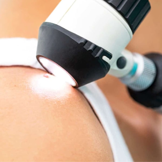
รูปภาพที่ 1 กล้อง Dermoscopy เพื่อประเมินสภาพผิว
(ที่มา: https://pallourasdermatology.com/wp-content/uploads/2022/01/New-Project-5-min.jpg)
เอกสารอ้างอิง
Fan L, He C, Jiang L, Bi Y, Dong Y, Jia Y. Brief analysis of causes of sensitive skin and advances in evaluation of anti-allergic activity of cosmetic products. Int J Cosmet Sci. 2016; 3 8(2): 120-7.
Berardesca E, Farage M, Maibach H. Sensitive skin: an overview. Int J Cosmet Sci. 2013; 35(1): 2-8.
Chen, B.; Tang, H.; Liu, Z.; Qiao, K.; Chen, X.; Liu, S.; Pan, N.; Chen, T.; Liu, Z. Mechanisms of Sensitive Skin and the Soothing Effects of Active Compounds: A Review. Cosmetics 2024, 11, 190.
Duarte I, Silveira JEPS, Hafner MFS, Toyota R, Pedroso DMM. Sensitive skin: review of an ascending concept. An Bras Dermatol. 2017; 92(4): 521-525.
Bodó, E.; Kovács, I.; Telek, A.; Varga, A.; Paus, R.; Kovács, L.; Bíró, T. Vanilloid receptor-1 (VR1) is widely expressed on various epithelial and mesenchymal cell types of human skin. J. Investig. Dermatol. 2004, 123, 410–413.
Itsekson A, Lazarov A, Cordoba M, Zeitune M, Abraham D, Seidman DS. Premenstrual syndrome and associated skin diseases related to hypersensitivity to female sex hormones. J Reprod Med. 2004; 49(3): 195-9.
Farage MA, Katsarou A, Maibach HI. Sensitive skin. Sensory, clinical, and physiological factors. In: Borel AO, Paye M, Maibach HI, editors. Handbook of Cosmetic Science and Technology. 4th ed. Boca Raton : CRC Press/Taylor & Francis Group, cop. 2014. p.59-69.
Dupont E, Gomez J, Bilodeau D. The two faces of skin erythema: sensitive skin and rosácea. Body Care Grooming, Protection e Hygiene. H&PC Today. 2013; 8: 12-14. (2013).
Lev-Tov H, Maibach HI. The sensitive skin syndrome. Indian J Dermatol. 2012;57(6):419-423.
Kim SJ, Lim SU, Won YH, An SS, Lee EY, Moon SJ, Kim J. The perception threshold measurement can be a useful tool for evaluation of sensitive skin. Int J Cosmet Sci. 2008;30:333-337.
Poomanee, W., Yaowiwat, N., Pattarachaidaecharuch, T., Leelapornpisid P. Optimized multiherbal combination and in vivo anti-skin aging potential: a randomized double blind placebo controlled study. Sci Rep 13, 5633 (2023).
Johansen JD, Aalto-Korte K, Agner T, Andersen KE, Bircher A, Bruze M, et al. European Society of Contact Dermatitis guideline for diagnostic patchtesting—Recommendations on best practice. Contact Dermatitis 73, 195–221 (2015).
Farage MA, Maibach HI, Andersen KE, Lachapelle JM, Kern P, Ryan C, et al. Historical perspective on the use of visual grading scales in evaluating skin irritation and sensitization. Contact. Derm.65(2), 65–75 (2011).
Kerscher, M, Buntrock, H. (2017). Treatments for Sensitive Skin. In: Honari G. Andersen RM. Maibach HI (ed.) Sensitive Skin Syndrome, Second Edition.
Nicholson, P.J., D. Dlewellyn and J.S. English. 2010. “Evidence-based guidelines for the prevention, identification and management of occupational contact dermatitis and urticaria.” Contact Dermatitis 63(4): 177-186.
Jeong S, Lee SH, Park BD, Wu Y, Man G, Man MQ. Comparison of the Efficacy of Atopalm® Multi-Lamellar Emulsion Cream and Physiogel® Intensive Cream in Improving Epidermal Permeability Barrier in Sensitive Skin. Dermatol Ther (Heidelb). 2016; 6(1): 47-56.
Varma, S.R.; Sivaprakasam, T.O.; Arumugam, I.; Dilip, N.; Raghuraman, M.; Pavan, K.B.; Rafiq, M.; Paramesh, R. In vitro anti-inflammatory and skin protective properties of Virgin coconut oil. J. Tradit. Complement. Med. 2019, 9, 5–14.
Qu, W.J. Application of hyaluronic acid in skin care. China Cosmet. Rev. 2020, 08, 94–97.
Lee, Y.I.; Lee, S.G.; Kim, J.; Choi, S.; Jung, I.; Lee, J.H. Proteoglycan Combined with Hyaluronic Acid and Hydrolyzed Collagen Restores the Skin Barrier in Mild Atopic Dermatitis and Dry, Eczema-Prone Skin: A Pilot Study. Int. J. Mol. Sci. 2021, 22, 10189.
Wahedi, H.M.; Jeong, M.; Chae, J.K.; Do, S.G.; Yoon, H.; Kim, S.Y. Aloesin from Aloe vera accelerates skin wound healing by modulating MAPK/Rho and Smad signaling pathways in vitro and in vivo. Phytomedicine 2017, 28, 19-26.
Kamatou GPP, Viljoen AM. A Review of the Application and Pharmacological Properties of α-Bisabolol and α-Bisabolol-Rich Oils. J Am Oil Chem Soc 2010; 87: 1–7.
Ren C, Ma Y, Wang Y, Luo D, Hong Y, Zhang X, et al. Palmitoylethanolamide-Incorporated Elastic Nano-Liposomes for Enhanced Transdermal Delivery and Anti-Inflammation. Pharmaceutics. 2024 Jun 29;16(7):876.
D’Agostino G., Russo R., Avagliano C., Cristiano C., Meli R., Calignano A. Palmitoylethanolamide Protects against the Amyloid-Β25-35-Induced Learning and Memory Impairment in Mice, an Experimental Model of Alzheimer Disease. Neuropsychopharmacology. 2012;37:1784–1792.
Ferreira MS, Sousa Lobo JM, Almeida IF. Sensitive skin: Active ingredients on the spotlight. Int J Cosmet Sci. 2022;44(1):56-73.
Boo, Y. C. (2021). Mechanistic Basis and Clinical Evidence for the Applications of Nicotinamide (Niacinamide) to Control Skin Aging and Pigmentation. Antioxidants, 10(8), 1315.
